









Abstract
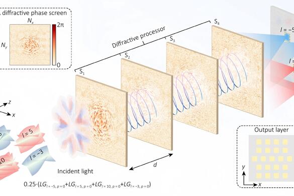
Beams carrying orbital angular momentum (OAM) have attracted considerable interest in high-capacity optical communication owing to their infinite-dimensional state space. Conventional methods for detecting OAM modes face significant limitations, including bulky systems, slow response times, and restricted detection ranges. Although deep learning algorithm have shown promise in mitigating some of these challenges, the characterization of mode distributions within mixed-mode OAM beams has received limited attention. We propose an all-optical, end-to-end approach for decomposing mixed-mode OAM beams and estimating their mode weights using diffractive deep neural network (D2NN). The network directly maps the incident optical field to outputs that both identify the constituent OAM modes and estimate their relative contributions. Numerical simulations demonstrate that the method can accurately recover the weights of up to 21 hybrid modes. Moreover, it maintains strong robustness under atmospheric perturbations, with the relative error remaining below 7%. This approach enables precise and efficient reconstruction of the OAM beams across varying numbers of modes, offering broad potential in multidimensional information encoding, quantum information processing, and optical computing.
Keywords
diffractive optical neural networks; orbital angular momentum; mixed-mode decomposition; mode-weight estimation
Acknowledgements
This research was supported by National Key Research and Development Program of China (No. 2021YFA1401003), and the National Natural Science Foundation of China (Nos. 62305345, U24A6010, 62222513).
Author contributions
L.-X.G. supervised the whole project. W.-L.J. performed the theoretical calculations and validation. W.-L.J. and W.-J.D. completed the initial structure and network construction. W.-L.J. and W.-J.D. contributed to the results analysis and data processing. W.-L.J. contributed to the data interpretation. W.-J.D. wrote the initial manuscript. W.-L.J., W.-J.D., H.-Y.L., and G.-Y.H. revised and edited the manuscript. All authors contributed to the discussions and preparation of the manuscript.
Competing interests
Xiangang Luo serves as an Editor for the Journal, and no other author has reported any competing interests.
References
[1] Al Ibrahim RH, Zheng SQ, Ng TK et al. Optical image rotation based on orbital angular momentum decomposition and combination. J Opt 24, 115605 (2022). DOI: 10.1088/2040-8986/ac8a02
[2] Li MM, Yan SH, Zhang YN et al. Orbital angular momentum in optical manipulations. J Opt 24, 114001 (2022). DOI: 10.1088/2040-8986/ac9192
[3] Andersen MF, Ryu C, Cladé P et al. Quantized rotation of atoms from photons with orbital angular momentum. Phys Rev Lett 97, 170406 (2006). DOI: 10.1103/PhysRevLett.97.170406
[4] Orlov S, Stanaitis K, Kizevičius P et al. Single-pixel terahertz imaging with enhanced edge detection using angular momentum of structured light. APL Photonics 10, 050805 (2025). DOI: 10.1063/5.0255550
[5] Li N, Zheng SL, He T et al. Metasurface-based dual-mode bright-field and spiral phase contrast THz imaging with enhanced focal depth. J Lightwave Technol 43, 4322–4330 (2025). DOI: 10.1109/JLT.2024.3521973
[6] Wang JK, Zhang WH, Qi QQ et al. Gradual edge enhancement in spiral phase contrast imaging with fractional vortex filters. Sci Rep 5, 15826 (2015). DOI: 10.1038/srep15826
Copyright
Open Access. © The Author(s). This article is licensed under a Creative Commons Attribution 4.0 International License. To view a copy of this license, visit http://creativecommons.org/licenses/by/4.0/.
Cite this Article
Wang LJ, Wang JD, Ha YL et al. Decomposition and mode-weight estimation of mixed-mode OAM beams via diffractive neural networks. Intell Opto-Electron 2, 250011 (2026).
Journals MORE
Articles MORE
Decomposition and mode-weight estimation of mixed-mode OAM beams via diffractive neural networks
Available online March 25, 2026 Intelligent Opto-Electronics
Advances in intelligent fiber-optic microfluidic-embedded technologies for empowered sensing performance: a review
Available online March 25, 2026 Intelligent Opto-Electronics
Abstract
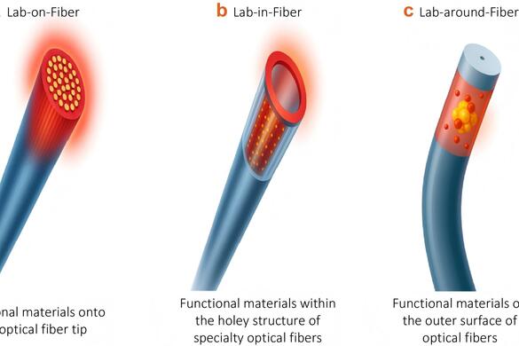
The convergence of Lab-on-Fiber (LoF) technology, microfluidics, and artificial intelligence (AI) is emerging as a new and powerful paradigm for next-generation intelligent sensing systems. Combining AI with LoF-microfluidic devices can cover the residual gap by enhancing precise microfluidic control, data analysis, adaptive calibration, and predictive sensing, thereby opening new pathways for intelligent, miniaturized, reliable, and multifunctional devices for biomedical sensing and environmental monitoring. Microfluidic technologies leverage high-precision and flow rate-controlled sample delivery, reagent optimization, and simple prototyping, which make them excellent for real-time sensing. LoF devices showcase unique light control at the nanoscale level and their integration onto microfluidic chips empowers signal-to-noise ratio and, ultimately, limit of detection in a controlled environment. Advances in materials science and engineering have allowed the realization of different types of nanostructures which are integrated onto fiber sensors whose performance can be optimally tuned to detect a variety of markers and molecules. However, open challenges still exist, such as scalability, reproducibility of results, detection of multiple targets, effective compensation of interfering parameters and fast data processing. Innovative AI-driven solutions and novel functional bio-/materials are being developed to overcome these barriers and possibly meet the future demands. A roadmap toward intelligent LoF-microfluidic platforms is finally envisioned.
Keywords
optical fiber sensors; biophotonics; microfluidics; artificial intelligence
Acknowledgements
F.C. acknowledges financial support under the National Recovery and Resilience Plan (NRRP), Mission 4, Component 2, Investment 1.1, Call for tender No. 1409 published on 14/09/2022 by the Italian Ministry of University and Research (MUR), funded by the European Union – NextGenerationEU – Project Title ‘‘Fiber optics sensors as a platform for cancer diagnosis and in vitro model testing (FOCAL)” – CUP B53D23024170001 – Grant Assignment Decree No. 1383 adopted on 01/09/2023 by the Italian MUR, and Project Title ‘‘Engineering Functional Metal Nanocluster-Protein Architectures for Bio(sensing and catalytic) applications (ProNano4Bio)” – B53D23013940006 – Grant Assignment Decree No. 958 adopted on 30/06/2023 by the Italian MUR. H.A. acknowledges financial support given by the Turkish Scientific and Technological Council (TÜBİTAK 1004-Regenerative and Restorative Medicine Research and Applications) under the grant numbers of 20AG003 and 20AG031, and by TÜBİTAK 1005-National New Ideas and Products under grant numbers of 123E013 and 225S581.
Author contributions
Shadab Dabagh and Francesco Chiavaioli conceived of the idea and designed the review. Rukmani Singh, Claudia Borri, Hamed Ghorbanpoor, Golara Ghorban Dordinejad, and Mahdi Bahadoran created and edited the figures. Shadab Dabagh led the manuscript writing – original draft. Shadab Dabagh, Ambra Giannetti, Francesco Baldini, Huseyin Avci, and Francesco Chiavaioli led the manuscript writing – review & editing. All authors participated in the review and discussion of the manuscript.
Competing interests
The authors declare no competing financial interests.
References
[1] Gupta BD, Shrivastav AM, Usha SP. Optical Sensors for Biomedical Diagnostics and Environmental Monitoring (CRC Press, Boca Raton, 2017).
[2] Sarabi MR, Jiang N, Ozturk E et al. Biomedical optical fibers. Lab Chip 21, 627–640 (2021). DOI: 10.1039/D0LC01155J
[3] Shah RY, Agrawal YK. Introduction to fiber optics: sensors for biomedical applications. Indian J Pharm Sci 73, 17–22 (2011).
[4] Wolfbeis OS. Fiber-optic chemical sensors and biosensors. Anal Chem 78, 3859–3874 (2006).
[5] Wang XD, Wolfbeis OS. Fiber-optic chemical sensors and biosensors (2013–2015). Anal Chem 88, 203–227 (2016). DOI: 10.1021/acs.analchem.5b04298
[6] Vaiano P, Carotenuto B, Pisco M et al. Lab on Fiber Technology for biological sensing applications. Laser Photonics Rev 10, 922–961 (2016).
Copyright
Open Access. © The Author(s). This article is licensed under a Creative Commons Attribution 4.0 International License. To view a copy of this license, visit http://creativecommons.org/licenses/by/4.0/.
Cite this Article
Dabagh S, Singh R, Borri C et al. Advances in intelligent fiber-optic microfluidic-embedded technologies for empowered sensing performance: a review. Intell Opto-Electron 2, 250015 (2026).
Polarization-multiplexed meta-neural networks for simultaneous imaging and all-optical classification
Available online March 25, 2026 Intelligent Opto-Electronics
Abstract
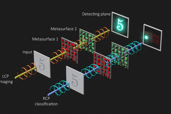
Deep learning has transformed perception and inference but remains constrained by memory–compute bottlenecks, latency, and energy costs. All-optical diffractive deep neural networks (D2NNs) alleviate these limitations by computing with light, yet most implementations trade image formation for direct classification, limiting downstream processing. Here we introduce a polarization-multiplexed meta-neural network (PMNN) that unifies imaging and classification within a single, static optical platform. The PMNN employs cascaded metasurfaces whose meta-atoms jointly harness geometric (Pancharatnam–Berry) and propagation phases to engineer distinct phase profiles for left- and right-circularly polarized (LCP and RCP) channels. This polarization contrast enables dual-channel functionality. Under LCP illumination, the system performs lens-like imaging, whereas under RCP illumination, it executes all-optical classification via diffractive routing to predefined detection regions. Built on a differentiable angular-spectrum forward model and trained end-to-end, the PMNN achieves 96.51% accuracy on handwritten-digit recognition while delivering an imaging mean squared error of 5.38×10⁻³, a peak signal-to-noise ratio of 22.70 dB, and a structural similarity index measure of 0.90. By coupling perception with inference without mechanical switching or electronic post-processing, the proposed approach enhances utility, reduces computational load, and offers a practical path toward compact, scalable, and energy-efficient optical intelligent systems.
Keywords
meta-device; metasurface; diffractive deep neural networks
Acknowledgements
This work is financed by the Guangdong Basic and Applied Basic Research Foundation [2025A1515011846], the National Science Foundation of China (NSFC) [62405254]; the Fundamental and Applied Fundamental Research Foundation Project of Guangdong Province [No. 2023A1515140108]; the University Grants Committee/Research Grants Council of the Hong Kong Special Administrative Region, China [CRF Project: C5031-22G; and GRF Project: CityU11310522; CityU11300123]; City University of Hong Kong [Project No. 9610628 and No. 7020142]; Guangdong and Hong Kong Universities '1+1+1' Joint Research Collaboration Scheme.
Author contributions
M.K.C. and J.C. conceived the idea for this work. M.K.C. supervised the research. J.C. is responsible for developing algorithms and implementing methods. M.Z. and Z.G. conceived the development of light field computing. X.L., J.C., and W.Z. built FDTD simulations. W.S. and Z. G. provided the high-speed computing hardware and guidance for the simulation work. J.C., X.L., and W.Z. perform data processing and analysis. All authors discussed the results and provided comments on the manuscript.
Competing interests
The authors declare no competing financial interests.
References
[1] He KM, Zhang XY, Ren SQ et al. Spatial pyramid pooling in deep convolutional networks for visual recognition. IEEE Trans Pattern Anal Mach Intell 37, 1904–1916 (2015). DOI: 10.1109/TPAMI.2015.2389824
[2] Guo YM et al. Adaptive optics based on machine learning: a review. Opto-Electron Adv 5, 200082 (2022).
[3] Dahl GE, Sainath TN, Hinton GE. Improving deep neural networks for LVCSR using rectified linear units and dropout. In 2013 IEEE International Conference on Acoustics, Speech and Signal Processing 8609–8613 (IEEE, 2013). http://doi.org/10.1109/ICASSP.2013.6639346.
[4] Collobert R, Weston J, Bottou L et al. Natural language processing (almost) from scratch. J Mach Learn Res 12, 2493–2537 (2011).
[5] Markram H. The blue brain project. Nat Rev Neurosci 7, 153–160 (2006). DOI: 10.1038/nrn1848
[6] Lin X, Rivenson Y, Yardimci NT et al. All-optical machine learning using diffractive deep neural networks. Science 361, 1004–1008 (2018). DOI: 10.1126/science.aat8084
Copyright
Open Access. © The Author(s). This article is licensed under a Creative Commons Attribution 4.0 International License. To view a copy of this license, visit http://creativecommons.org/licenses/by/4.0/.
Cite this Article
Cheng JL, Li X, Zhu WJ et al. Polarization-multiplexed meta-neural networks for simultaneous imaging and all-optical classification. Intell Opto-Electron 2, 250017 (2026).
Research progress in integrated optics for optical coherence tomography
Available online March 25, 2026 Intelligent Opto-Electronics
Abstract
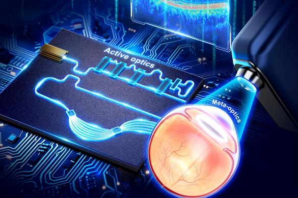
Optical coherence tomography (OCT) is a noninvasive biomedical imaging technique that exploits the back-reflection and scattering properties of tissue without the need for exogenous contrast agents. It enables high-resolution, in situ visualization of tissue microstructures and pathological features, without requiring specimen removal or processing. OCT offers cross-sectional and three-dimensional imaging of biological tissues with micron-level resolution and millimeter-scale depths. When integrated with an endoscope, OCT becomes a powerful and versatile imaging tool in the medical field. However, conventional OCT systems face several limitations, including a bulky system volume of approximately 1 m3, high costs of around $100,000, operational complexity, limited portability, and the need for precise optical alignment, which demands substantial manpower and time. To overcome these challenges, integrated optics has emerged as a promising solution. In recent years, significant advances have been made in developing OCT systems based on integrated photonics. All the integrated-optics devices, such as light sources, isolators, couplers, circulators, detectors, and active optical components, can be exploited for endoscopic and non-endoscopic OCT systems and enable more compact designs and implementations. This review provides a comprehensive overview of these advancements. We first summarize the fundamental principles and imaging properties of OCT, along with the design and functionality of OCT endoscopic probes. We then examine the recent progress in on-chip OCT systems, focusing on system optimization and the implementation of integrated photonic technologies. Finally, we discuss the current challenges, including the full integration of optical components onto a single chip, and explore prospects of integrated-optics based OCT endoscopy, particularly the integration of AI-powered intelligent diagnostics to enhance real-time clinical decision-making and expand the applications of OCT in personalized medicine.
Keywords
Acknowledgements
This work was financially supported by the National Natural Science Foundation of China under Grant (62205306); Fudan University through the Research Initiation Project (IDH2323007Y, IDH2323008Y, IDH2323010Y) and the National Natural Science Foundation of China.Q. Q. Song thanks the financial support from the National Natural Science Foundation of China under Grant (62205306). G. Li thanks the financial support from Fudan University through the Research Initiation Project (IDH2323007Y, IDH2323008Y, IDH2323010Y) and the National Natural Science Foundation of China.
Author contributions
Writing-original draft preparation: Q. Q. S. Writing-review and editing: Q.Q. S, G. Q. L. Review, editing and discussion: D. Z, Q. Q. S, G. Q. L, Z. D. All authors read and approved the final manuscript.
Competing interests
The authors declare no competing financial interests.
References
[1] Huang D, Swanson EA, Lin CP et al. Optical coherence tomography. Science 254, 1178–1181 (1991). DOI: 10.1126/science.1957169
[2] Drexler W, Morgner U, Ghanta RK et al. Ultrahigh-resolution ophthalmic optical coherence tomography. Nat Med 7, 502–507 (2001). DOI: 10.1038/86589
[3] Bouma BE, Tearney GJ, Compton CC et al. High-resolution imaging of the human esophagus and stomach in vivo using optical coherence tomography. Gastrointest Endosc 51, 467–474 (2000). DOI: 10.1016/S0016-5107(00)70449-4
[4] Tearney GJ, Brezinski ME, Bouma BE et al. In vivo endoscopic optical biopsy with optical coherence tomography. Science 276, 2037–2039 (1997). DOI: 10.1126/science.276.5321.2037
[5] Tearney GJ, Brezinski ME, Boppart SA et al. Catheter-based optical imaging of a human coronary artery. Circulation 94, 3013 (1996). DOI: 10.1161/01.CIR.94.11.3013
[6] Hanna N, Saltzman D, Mukai D et al. Two-dimensional and 3-dimensional optical coherence tomographic imaging of the airway, lung, and pleura. J Thorac Cardiov Surg 129, 615–622 (2005). DOI: 10.1016/j.jtcvs.2004.10.022
Copyright
Open Access. © The Author(s). This article is licensed under a Creative Commons Attribution 4.0 International License. To view a copy of this license, visit http://creativecommons.org/licenses/by/4.0/.
Cite this Article
Song QQ, Zhang DW, Ding ZH et al. Research progress in integrated optics for optical coherence tomography. Intell Opto-Electron 2, 250009 (2026).
optical coherence tomograpghy; imaging; endoscope; integrated optics